Protein stabilizers are essential for maintaining the structure and function of protein-based drugs, which are sensitive to environmental factors like temperature, oxidation, and mechanical stress. These stabilizers, such as amino acids, polymers, and surfactants, help prevent protein degradation, ensuring drug safety and efficacy.
Testing stabilizers involves analyzing protein stability under stress conditions like heat, light, and pH changes. Common methods include spectroscopy (e.g., Circular Dichroism), chromatography (e.g., Size Exclusion Chromatography), and thermal analysis (e.g., Differential Scanning Calorimetry). These tests identify degradation pathways, such as aggregation and oxidation, which can reduce drug potency or trigger immune responses.
Regulatory guidelines like ICH Q5C and Q2(R2) require validated, stability-indicating methods to ensure biopharmaceuticals meet quality standards. Proper documentation and high-quality reagents are crucial for compliance. By addressing these challenges, companies can extend shelf life and maintain the therapeutic value of protein-based drugs.
From Molecule to Formulation: A Systematic Approach to Assess Biologics Developability
sbb-itb-aa4586a
Protein Degradation Pathways and Their Effects
Proteins can break down in two main ways: chemically and physically. Chemical degradation involves changes to covalent bonds through processes like oxidation, deamidation, disulfide scrambling, and hydrolysis [3][5]. Physical degradation, on the other hand, disrupts the structural integrity of proteins – breaking down their secondary, tertiary, or quaternary structures, which can lead to unfolding, denaturation, aggregation, and precipitation [3][5]. Often, these two types of degradation are interconnected; for instance, oxidation can set off a chain reaction that results in protein aggregation [3][5].
The impact of protein degradation goes beyond just quality control. Degraded proteins may lose their biological activity, either partially or entirely. For example, the deamidation of growth-hormone-releasing factor can reduce its bioactivity by as much as 500-fold, depending on the isomer formed [3]. Beyond reduced effectiveness, degraded proteins can also pose safety risks. Aggregates, in particular, can provoke immune responses by being recognized as "foreign", which may lead to the production of anti-drug antibodies and unwanted immunogenicity [3][5].
Aggregation and Its Challenges
Protein aggregation typically begins when structural changes, like denaturation, cause proteins to self-associate. Initially, these interactions are reversible, but they can progress into irreversible complexes that resist separation. As more monomers join, these aggregates grow, forming soluble complexes that can eventually precipitate into insoluble particles. Several factors drive this process, including mechanisms like 3D domain swapping (where partially unfolded monomers exchange structural domains), the formation of interchain salt bridges, and oxidative stress that disrupts proper protein folding.
Aggregation can drastically reduce a product’s potency. A striking example occurred in 2011 when bevacizumab (Avastin) lost half of its active IgG concentration after being repackaged at a compounding pharmacy. Handling and mechanical manipulation at such facilities have been shown to increase aggregation levels by up to tenfold compared to the original product [3].
Experts Andrea Hawe, John F. Carpenter, and Wim Jiskoot have emphasized the severity of this issue:
Unwanted immunogenicity often has severe implications for the efficacy and safety of biopharmaceuticals. Among the several factors playing a role in immunogenicity, the presence of degraded protein in therapeutic formulations is considered to be a major risk [5].
Chemical degradation, especially oxidation, further exacerbates the stability challenges faced by protein therapeutics.
Oxidation and Stability Concerns
Oxidation damages proteins by modifying specific amino acids, particularly those containing sulfur atoms or aromatic rings, such as methionine, cysteine, histidine, tryptophan, and tyrosine [3]. Common triggers include light exposure, metal ions, peroxides, and oxygen in the headspace of vials. The pH of the environment also plays a critical role. Alkaline conditions can accelerate methionine and cysteine oxidation by deprotonating thiol groups, while acidic conditions can promote photosensitized reactions affecting methionine and tryptophan [3]. For instance, oxygen in the headspace of multidose vials caused a 50% loss of tuberculin protein potency within four months [3].
To mitigate oxidation, inert gases like nitrogen or argon can replace headspace oxygen [3]. Another approach involves adding "sacrificial targets", such as L-methionine or ascorbic acid, which absorb oxidizing agents before they can harm the protein. Chelating agents like EDTA or citrate can also help by binding transition metal ions and preventing metal-catalyzed oxidation [3].
Testing stabilizers under controlled oxidative conditions is a critical step. This process evaluates how well a formulation maintains potency, structural integrity, and resistance to aggregation over time, often through the use of surfactants to prevent protein aggregation.
Thermal and Chemical Instabilities
Temperature and pH fluctuations can also destabilize proteins. High temperatures cause thermal denaturation, where a protein’s secondary and tertiary structures unfold cooperatively [6]. Freezing presents another challenge: ice crystal formation concentrates proteins and excipients, leading to pH shifts and a "salting out" effect. For example, thyroid-stimulating hormone lost over 40% of its potency within 90 days when stored at –20°C (about –4°F), even though it remained stable at –80°C (about –112°F) or 4°C (about 39°F) [3].
Chemical stressors like pH shifts further accelerate degradation. Keeping formulations within a pH range of 3–5 can minimize peptide deamidation [3]. Buffer selection is equally important, as stability can vary depending on the buffer used. For instance, a protein may remain more stable in sodium phosphate at pH 7.0 compared to HEPES or Tris at the same pH [6]. Amine buffers like Tris and histidine are particularly sensitive to temperature changes, which can cause pH shifts that promote deamidation and oxidation [3]. In one study, reducing the dielectric strength of the medium from 80 (water) to 35 (using a PVP/glycerol/water mixture) decreased peptide deamidation rates by sixfold [3].
To address these challenges, top stabilizers for protein-based biopharmaceuticals should be evaluated under realistic temperature and pH conditions. Rigorous testing and well-defined stress protocols are essential to ensure protein stability in therapeutic formulations.
This content is for informational purposes only. Consult official regulations and qualified professionals before making sourcing or formulation decisions.
Analytical Techniques for Testing Protein Stabilizers
Analytical Methods for Protein Stabilizer Testing in Biopharmaceuticals
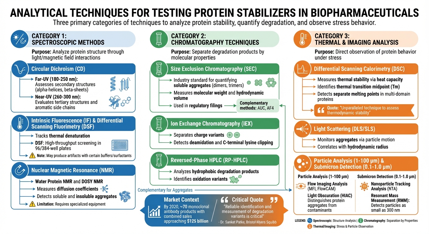
Evaluating the effectiveness of protein stabilizers involves analyzing protein structure, aggregation, and bioactivity. This is achieved through three primary types of analytical methods: spectroscopic techniques, chromatography, and thermal or imaging analysis. Below, we’ll break down these approaches and their role in confirming stabilizer performance.
Spectroscopic Methods
Spectroscopic techniques explore protein structure by examining how light or magnetic fields interact with proteins. Circular Dichroism (CD), for instance, uses polarized light to assess secondary structures like alpha-helices and beta-sheets in the far-UV range (180–250 nm). Near-UV CD (260–300 nm), on the other hand, provides insights into tertiary structures and aromatic side chain transitions [8].
Intrinsic Fluorescence (IF) and Differential Scanning Fluorimetry (DSF) are also key tools. These methods track thermal denaturation by observing changes in the protein’s environment or dye binding as the protein unfolds [7][8]. DSF is particularly useful for high-throughput screening, as it works with small sample volumes in 96- or 384-well plates [8]. However, fluorescence assays can sometimes produce artifacts due to specific buffers or surfactants, making it essential to validate results with calorimetric techniques [7].
Another powerful tool is Nuclear Magnetic Resonance (NMR) spectroscopy. Methods like Water Protein NMR (_w_NMR) and Diffusion Ordered Spectroscopy (DOSY) NMR measure diffusion coefficients, allowing scientists to detect both soluble and insoluble aggregates [2]. While highly effective, these techniques require specialized equipment and expertise, making them less accessible than other methods.
Chromatography Techniques
Chromatography focuses on separating degradation products based on molecular properties. Size Exclusion Chromatography (SEC) is widely used in the industry to quantify soluble aggregates, such as dimers and trimers [2]. It’s a staple in regulatory filings for protein therapeutics because it reliably measures molecular weight and hydrodynamic volume. However, SEC isn’t perfect – some aggregates may interact with the column matrix and escape detection. For this reason, regulatory agencies often recommend using complementary methods like Analytical Ultracentrifugation (AUC) or Asymmetrical Flow Field-Flow Fractionation (AF4) to confirm findings [2].
Ion Exchange Chromatography (IEX) is another critical technique, separating charge variants caused by chemical changes like deamidation or C-terminal lysine clipping [2]. This method helps evaluate how stabilizers prevent chemical modifications that alter a protein’s surface charge. To complement IEX, Reversed-Phase HPLC (RP-HPLC) is used to analyze hydrophobic degradation products and oxidation variants. Dr. Sanket Patke from Bristol-Myers Squibb underscores the importance of these methods:
Reliable identification and measurement of degradation variants is critical [2].
Thermal and Imaging Analysis
Thermal and imaging analyses offer a direct look at how proteins behave under stress, supplementing spectroscopic and chromatographic data. Differential Scanning Calorimetry (DSC) is a widely trusted method for measuring thermal stability [7]. By tracking heat capacity, DSC identifies the thermal transition midpoint ($T_m$) – the temperature where half the protein is unfolded. A higher $T_m$ suggests greater stability and a reduced risk of aggregation [7]. DSC is particularly valuable for multi-domain proteins like monoclonal antibodies, as it can detect separate melting points for different domains (e.g., CH2, Fab, CH3 regions), which spectroscopic methods might overlook [7]. As noted by Gokarn and colleagues:
DSC remains as an unparalleled technique to assess the thermodynamic stability of proteins in a given buffer condition [7].
Dynamic Light Scattering (DLS) and Static Light Scattering (SLS) are commonly used to monitor aggregates by correlating particle motion with hydrodynamic radius [8]. For larger particles (1 to 100 µm), Flow Imaging Analysis (using tools like MFI or FlowCAM) and Light Obscuration (HIAC) are standard methods [2][7]. Flow imaging provides visual data on particle size, shape, and transparency, helping distinguish protein aggregates from contaminants like silicone oil droplets or air bubbles [2]. Meanwhile, Resonant Mass Measurement (RMM) can detect particles as small as 300 nm, differentiating between negatively buoyant protein particles and positively buoyant silicone oil [2].
With increasing regulatory attention on submicron particles (0.1–1.0 µm), techniques like Nanoparticle Tracking Analysis (NTA) and RMM have gained prominence [2]. NTA offers more precise differentiation of particle subpopulations by tracking individual particles under Brownian motion, a step up from bulk measurements provided by DLS [2]. By 2020, the global market for monoclonal antibody products was projected to include around 70 products, with combined sales nearing $125 billion [2]. This scale underscores the need for rigorous analytical validation at every stage of development.
This content is for informational purposes only. Consult official regulations and qualified professionals before making sourcing or formulation decisions.
Stability Study Design and Stress Testing Protocols
Designing protein stabilizer studies requires meticulous planning to ensure formulations can endure the stresses of storage and handling. Proteins are particularly sensitive to environmental changes, and their biological activity relies on maintaining a precise molecular structure, held together by both covalent and noncovalent interactions [1]. Stability studies are essential to evaluate how well a protein retains its structural integrity under stress, forming the basis for defining specific test parameters.
Developing Test Parameters
The first step in designing a stability study is identifying the environmental stressors the formulation may face. Regulatory guidelines emphasize evaluating multiple factors, such as temperature, oxidation, light exposure, ionic strength, and mechanical stress [9]. For instance, the ICH Q5C guideline outlines a regulatory framework for biotechnological product stability testing, highlighting the need to tailor studies to the unique properties of each protein [9]. For example, a monoclonal antibody stored at 2°C to 8°C (35°F to 46°F) might still degrade if exposed to repeated freeze–thaw cycles or changes in buffer ionic strength during storage.
In addition to real-time storage conditions, accelerated aging studies (e.g., 25°C, 60% RH) should be included to predict long-term stability. However, temperature stress alone is insufficient. Protocols must also simulate mechanical stresses, like agitation or pumping, and assess stabilizer performance under varying salt concentrations or pH levels. For formulations administered via pre-filled syringes, shear stress testing is critical to replicate the forces experienced during injection.
Data Interpretation and Decision-Making
Once test parameters are established, accurate data interpretation becomes crucial for validating stabilizer effectiveness. The primary goal isn’t just to confirm chemical purity but to ensure the protein maintains its biological activity throughout its storage life. Since protein function depends on maintaining its molecular conformation, even minor structural changes can indicate early stabilizer failure, often before chemical degradation is detectable.
To draw meaningful conclusions, data from multiple analytical methods should be correlated. For instance, if differential scanning calorimetry reveals a lower thermal transition midpoint (Tₘ), size-exclusion chromatography shows no soluble aggregates, and circular dichroism spectroscopy detects secondary structure changes, while reverse-phase HPLC finds no signs of oxidation, it could signal that the stabilizer is failing to protect noncovalent interactions despite an intact primary sequence. The focus should remain on whether the stabilizer preserves the protein’s functional state under the identified environmental stressors [9]. If biological activity falls below acceptable thresholds – even with an intact primary sequence – the stabilizer has not performed adequately.
To ensure reliable results, laboratories must use high-quality and consistent chemical reagents. Collaborating with a dependable supplier, such as Allan Chemical Corporation (https://allanchems.com), can provide access to regulatory-compliant materials that support robust and reliable stability testing.
This content is for informational purposes only. Always consult official regulations and qualified professionals when making sourcing or formulation decisions.
Regulatory Requirements for Protein Stabilizer Validation
Regulatory validation plays a critical role in confirming that protein stabilizers perform as intended throughout a product’s lifecycle. This process, grounded in well-established analytical techniques, ensures that biopharmaceuticals remain both safe and effective over time. Adhering to frameworks set by the FDA and ICH is essential for bringing protein-based therapeutics to market and maintaining compliance throughout their shelf life.
Meeting Regulatory Standards
ICH Q5C is the cornerstone guideline for biological products, emphasizing the need for specialized stability testing for proteins and polypeptides. As the guideline notes:
Biotechnological/biological products have distinguishing characteristics to which consideration should be given in any well-defined testing program designed to confirm their stability during the intended storage period [9].
This underscores the importance of testing programs that verify stabilizers maintain molecular structure and biological activity under storage conditions.
The revised ICH Q2(R2) (March 2024) provides additional clarity, requiring stability-indicating analytical methods that demonstrate specificity, accuracy, precision, and a suitable reportable range [10]. For protein stabilizers, these methods must identify changes in quality attributes, such as aggregation or degradation, during storage. According to ICH Q2(R2):
A validated quantitative analytical procedure that can detect changes in relevant quality attributes of a product during storage is considered to be stability indicating [10].
For impurity testing, the reportable range must span from the reporting threshold to 120% of the specification limit [10]. Complementing Q2(R2), ICH Q14 provides a science-driven framework for developing analytical procedures, ensuring methods are purpose-built from the outset.
To confirm specificity in stability tests, companies must use stressed samples – exposed to conditions like heat, oxidation, or light – to prove the method can distinguish intact proteins from degraded forms. This ensures that any stabilizer failure is detected accurately and reliably [10].
Once analytical methods are validated, thorough documentation becomes essential for maintaining compliance.
Documentation and Compliance
Comprehensive documentation is a cornerstone of regulatory compliance during the validation process. Before initiating validation studies, companies must create a formal validation protocol that outlines the study’s purpose, the performance characteristics to be tested, and predefined acceptance criteria [10]. This protocol acts as a blueprint, demonstrating to regulators that the validation effort is systematic and well-planned.
After the study concludes, a validation report must summarize the findings, compare results against the protocol’s criteria, and determine whether the method is fit for its intended purpose [10]. All supporting data, including calculation methodologies, must be included in the regulatory application, following the ICH M4Q(R1) format.
For reference materials used in validation, documented identity and purity are critical to ensure results are based on well-characterized standards [10]. Multivariate analytical procedures, such as near-infrared (NIR) or Raman spectroscopy, require additional documentation for both model development and independent validation phases using challenge samples [10].
Validation doesn’t end with initial approval. Any procedural changes demand risk-based revalidation, and laboratories transferring methods must confirm consistency through comparative analyses. System suitability tests should also be integrated into the analytical procedure to ensure consistent performance during routine use [10].
For companies conducting validation studies, sourcing high-quality chemical reagents is vital. Reliable suppliers, such as Allan Chemical Corporation (https://allanchems.com), can provide compendial-grade materials that meet regulatory standards, supporting consistent and compliant testing programs.
This content is for informational purposes only. Always consult official regulations and qualified professionals before making sourcing or formulation decisions.
Conclusion
Thorough testing of biopharmaceuticals requires a strategic combination of advanced analytics, stress testing, and strict compliance with regulatory standards. Nadine Ritter, President of the CASSS Board of Directors, underscores this point:
The technical challenge is to use a complete set of orthogonal analytics that can detect potential physical and functional changes in a product if it should degrade. [4]
Biological products, unlike small molecules, exhibit intricate degradation patterns. This makes real-time stability data indispensable for accurately predicting shelf life. A well-rounded approach to testing helps navigate the regulatory and market complexities tied to these products.
Forced degradation studies – using heat, light, oxidation, and agitation – play a critical role in validating stability-indicating methods. [4] These studies not only identify effective stabilizers, such as Trehalose, Sucrose, L-Histidine, and Polysorbates 20 and 80, but also ensure analytical methods can detect degradation through multiple pathways. The stakes are high: protein degradants can lead to immunogenic responses, posing serious safety risks if stability data are unreliable. [4]
Regulatory validation builds on these analytical efforts to ensure consistent protein quality. Frameworks like ICH Q5C and Q6B mandate compliance and require excipients that meet USP-NF standards. Even trace impurities, such as peroxides in low-quality polysorbates, can trigger protein oxidation, jeopardizing carefully designed formulations. [3] Reliable suppliers, such as Allan Chemical Corporation (https://allanchems.com), provide compendial-grade stabilizers with the necessary documentation to support rigorous testing programs.
By addressing these testing challenges, companies are better equipped to meet the growing demand for safe and effective biopharmaceuticals. Peptide and protein drugs now represent about 25% of the global pharmaceutical market, driving the need for reliable stabilizer testing. [11] Companies that adopt Quality by Design (QbD) principles, track operational metrics like invalid rates, and maintain meticulous documentation will be well-positioned to ensure their products remain safe and effective throughout their intended shelf life.
This content is for informational purposes only. Consult official regulations and qualified professionals before making sourcing or formulation decisions.
FAQs
What factors influence protein stability in biopharmaceuticals?
Protein stability in biopharmaceuticals depends on a variety of factors, including external conditions like temperature, light, oxidation, ionic strength, and mechanical stress (e.g., shear forces). These factors can trigger degradation processes such as denaturation, aggregation, or chemical modifications, which may reduce the protein’s effectiveness or increase the risk of immune reactions.
The formulation itself also plays a key role. Variables like pH, excipients, and storage conditions must be carefully managed to maintain the protein’s structure and biological function. Properly optimizing these factors can extend shelf life and ensure consistent performance.
To keep track of stability, advanced tools like thermal unfolding assays are commonly employed during both development and storage. These methods provide critical insights into how proteins degrade and help verify their structural integrity. Managing these stability factors is crucial to ensure biopharmaceuticals remain safe and effective.
What role do regulatory guidelines play in protein stabilizer testing?
Regulatory guidelines are essential in shaping how protein stabilizer testing is conducted and documented, setting clear standards for stability studies. In the United States, the FDA adheres to frameworks like ICH Q5C, which focus on preserving the molecular structure and biological activity of proteins and peptides. These guidelines ensure that stability testing addresses key environmental factors – such as temperature, light, oxidation, and shear stress – that could compromise a product’s integrity and effectiveness.
Additionally, regulatory bodies mandate the use of reliable analytical methods to evaluate stability. These include tests designed to measure biological activity and confirm molecular conformation. By establishing these standards, agencies aim to ensure that storage conditions maintain product quality throughout its shelf life, safeguarding both patient safety and the product’s therapeutic performance.
What are the best methods for detecting protein degradation in biopharmaceuticals?
Detecting protein degradation in biopharmaceuticals relies heavily on techniques like chromatography, including size-exclusion and reverse-phase methods, as well as mass spectrometry. These tools are particularly effective at pinpointing issues such as chemical or physical instability, aggregation, and the formation of particulates.
Beyond these, biophysical methods – such as thermal shift assays and high-throughput screening – offer key insights into protein stability and aggregation tendencies. Together, these approaches play a crucial role in maintaining the quality and effectiveness of biopharmaceutical products.


Comments are closed